
Yuanyuan Li (CSIF) : Highly-multiplexed tonsil tissue imaged with Phenocycler Fusion
Main content start
The Cell Sciences Imaging Facility (CSIF) is a Beckman Center and Stanford Cancer Institute supported university service center that provides high resolution, state-of-the-art light and electron microscopy technologies for imaging and analyzing the molecular and structural organization of cells, tissue and bioengineered materials. The CSIF operates two sites at Stanford University: the SOM Beckman Center CSIF and the SOE Shriram Center CSIF. These sites are open to all members of the Stanford community as well as to external academic and industry researchers (with approval of Jon Mulholland, CSIF Director).

Confocal. Judith Maria Kraiczy, Human Fallopian Tube Cells

Highly-Multiplexed TMA - Phenocycler (Yuanyuan Li, CSIF)
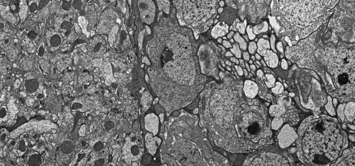
Transmission Electron Microscopy. Fly Brain 1200x (John Perrino, CSIF)
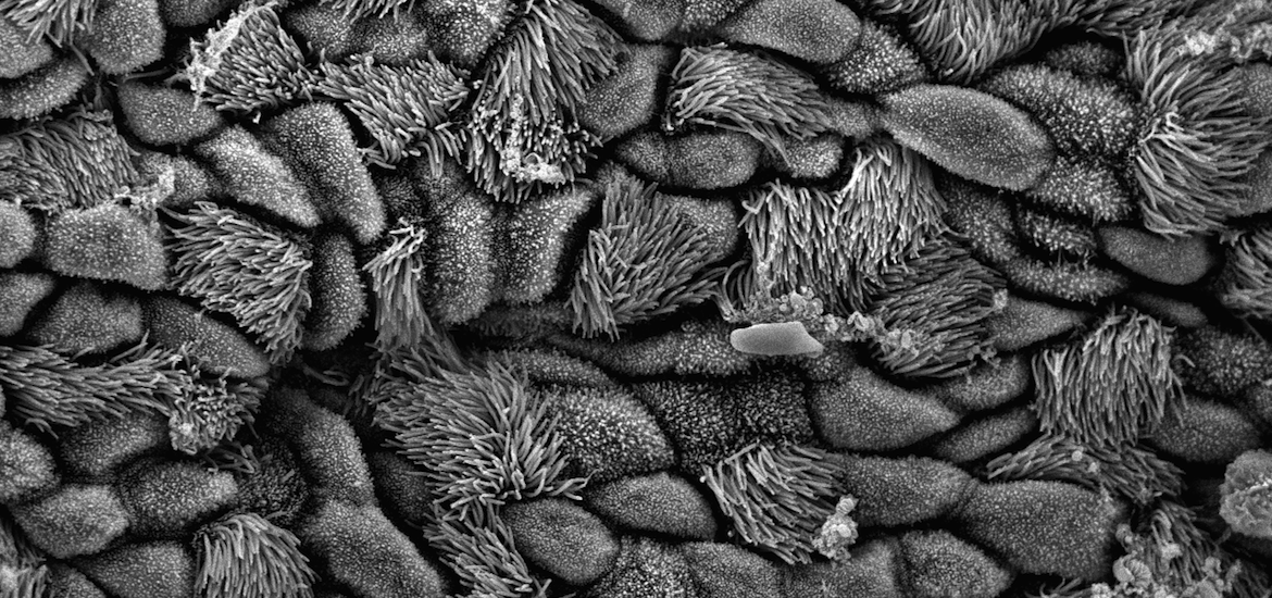
Scanning Electron Microscopy. Cilia mouse trachea
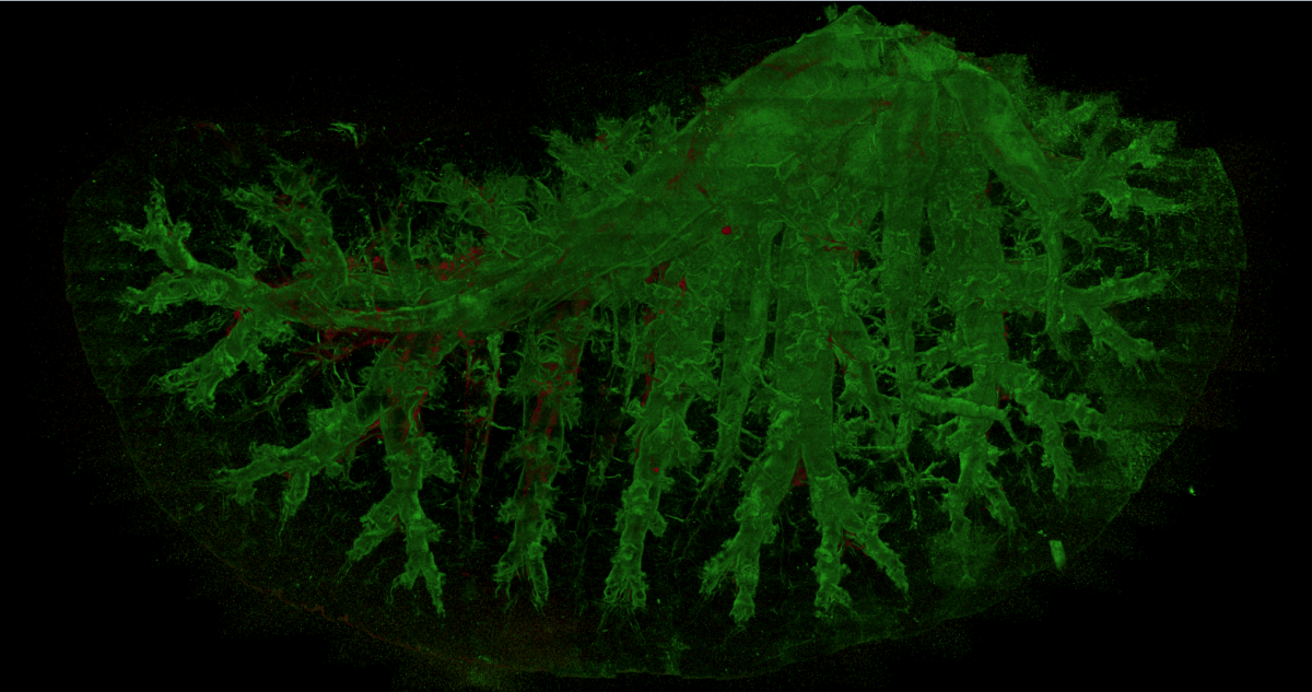
Tiled Confocal Image. Mouse lung.
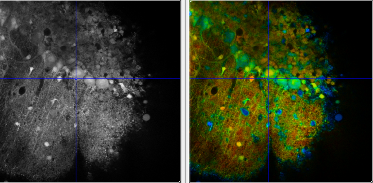
Fluorescence Life Time Microscopy (FLIM). FLIM image of mouse cerebellum purkinje cells
Technologies Offered
Light Microscopy
- Confocal microscopy (scanning and spinning-disk)
- Two-photon microscopy
- Wide-field fluorescence microscopy
- Digital deconvolution
- Transmitted-light imaging (phase, DIC, histology)
- High-content screening (Confocal and Wide-field)
- Super-resolution imaging (SIM and AiryScan)
- Cell surface imaging with <100 nM z-resolution (TIRF)
- Specialized microscopy (FRAP, FRET, FLIP...)
- Fluorescence lifetime imaging microscopy (FLIM)
- Lattice Light Sheet Microscopy (LLSM)
- Light Sheet Microscopy (LSM)
Spatial Proteomics - Highly Multiplexed Imaging
- Array Tomography
- Akoya PhenoCycler Fusion and PhenoCycler (CODEX) services (50+ biomarkers)
Atomic Force Microscopy
- Bruker BioScope Resolve integrated onto a Zeiss LSM900/AiryScan2
Location

Events
-
Imaris 10.1.1 updated, Adobe Creative Cloud Available!
AI segmentation in Imaris, Latest Adobe product Access-